Intra-Skeletal, Intra-Specific, and Inter-Specific Osteohistological Variability
Determining Best Practices for of Studies Growth Inference & Body Size Evolution
Osteohistological techniques are becoming more and more common in studies examining the growth, age, and physiology of extinct vertebrates, Unfortunately, the variability in the proxies used for these inferences remains largely unquantified, and considerable debate exists regarding the optimal skeletal elements that should be used in these investigations. This project, which began through side-projects during my MSc and PhD, has expanded to form my primary postdoctoral research. Broadly, the goals of this project are to quantify the variability that exists in osteohistological features (such as growth mark count, growth zone area/spacing, vascularity and tissue structure, and osteocyte lacunar density/size/shape) at multiple scales (including within a single bone, between different bones of the same individual skeleton, between individuals of the same species, and between species), and use those data to test hypotheses of body-size evolution in non-avian dinosaurs.
|
While this project is ongoing, earlier components of the project have been published, and are detailed here:
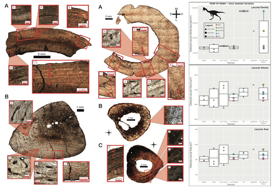
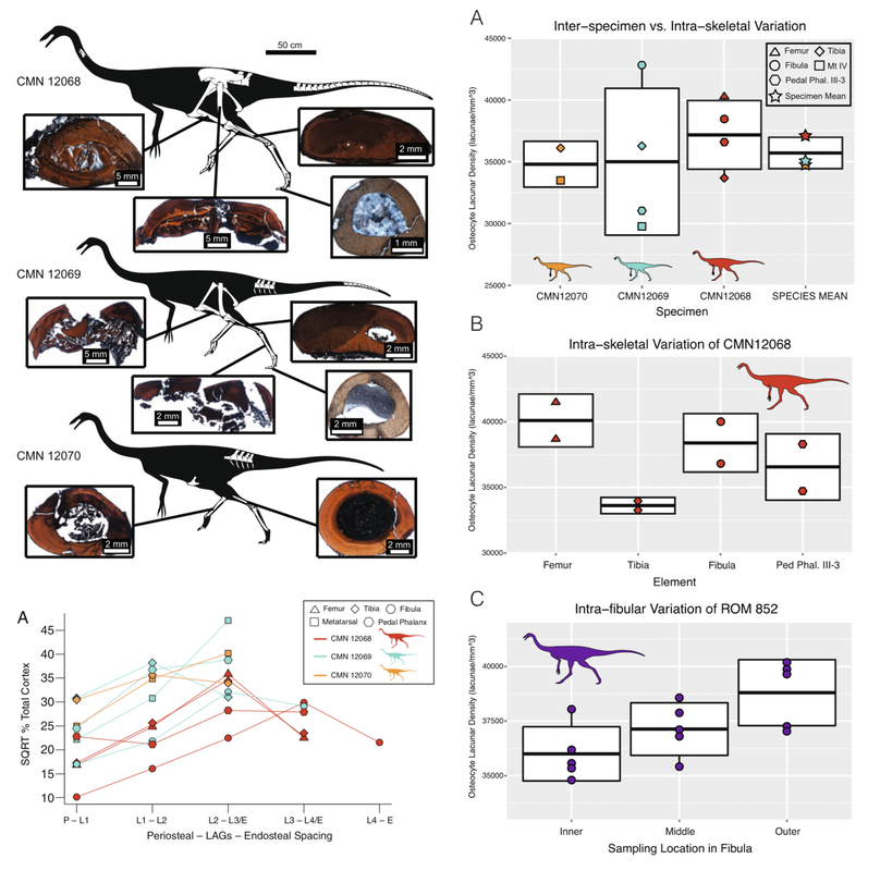
In this paper we described the osteohistology of multiple hind limb elements from a series of ornithomimids and used them as a case study to assess the range of intra-specific and intra-individual variation present in LAG spacing and osteocyte lacunar density. Different patterns of LAG spacing were found between upper and lower hind limb bones, suggesting that direct comparisons of these elements may be misleading, and that LAG spacing is not a reliable proxy for individual growth rates. Osteocyte lacunar density varied more between individual bone elements than between average individual values, suggesting that the choice of sampled element can greatly influence the result, and care should be taken to not bias interpretations of the physiology of fossil tetrapods.
This research was published in BMC Evolutionary Biology, was an Editor’s Pick and a BMC Series Highlight for November 2014, and our figures were recognized as among the top images from the journal for 2014..
We continued this work with similar examinations of the caenagnathid Anzu, finding many similar patterns as we had observed in ornithomimids. Our work on Anzu also has cautionary implications for the relatively common practice of naming species or assigning specimens to species based on size in this group, without ontogenetic/histological assessments. This work has been reviewed and accepted in Papers in Palaeontology, and should be published before the end of 2019.
Above: comparative osteohistology of Anzu, from Cullen et al Accepted, Papers in Palaeontology
|
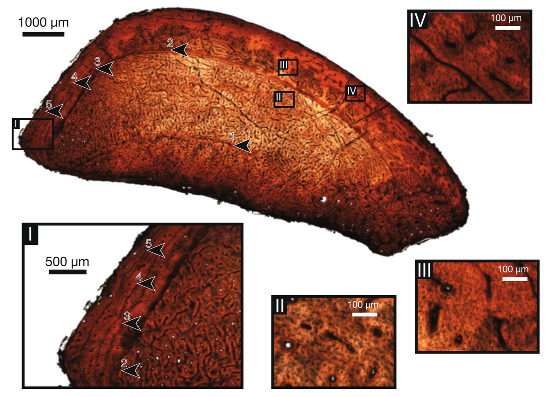
Above: bone microstructure of an ornithomimid fibula; Below: comparative osteohistology in ornithomimids. From Cullen et al 2014b
|
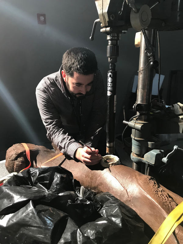
(preparing to core sample a large theropod femur in 2018, photo courtesy of D. Evans)
|
The next stage of this project formed the core of my postdoctoral research at the Field Museum of Natural History.
Alongside with Pete Makovicky, I continued my research into osteohistological variability, with a greater focus on intra-specific and intra-skeletal variation between weight-bearing and non weight-bearing elements. These investigations were designed to enhance our ability to perform accurate growth and age reconstructions of fossil organisms, and better understand which bones may preserve the best growth record in particular groups and at particular parts of the ontogenetic series. This will facilitate better growth curve reconstruction, and allow testing of hypotheses of the independent evolution of large body size in multiple dinosaur clades, with a particular emphasis on theropod 'gigantism'. Most of this is currently in progress or under peer-review. Stay tuned for updates. 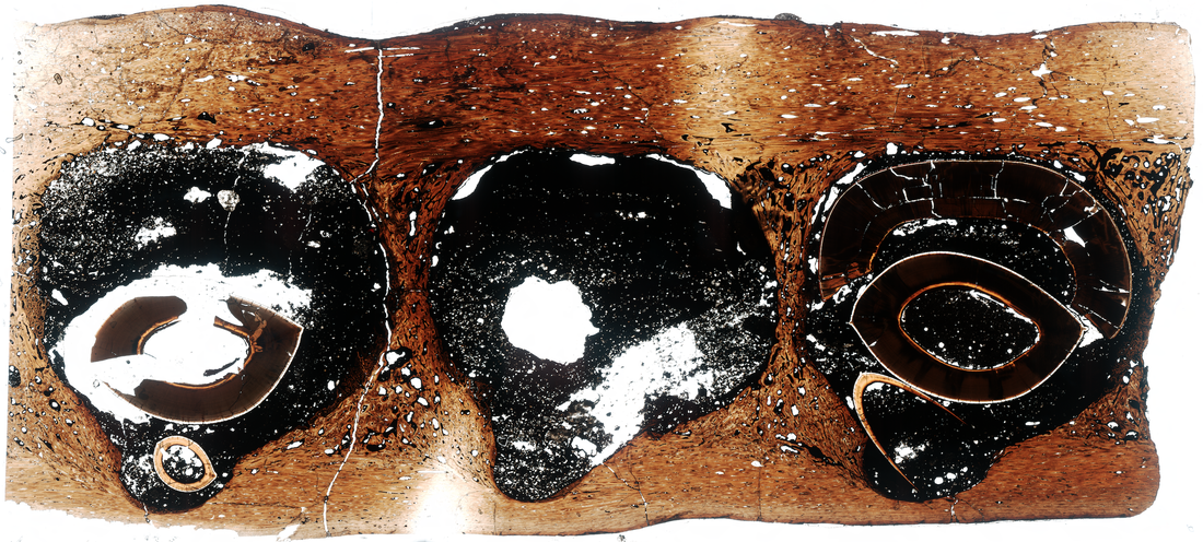
(thin-section of a Gorgosaurus jaw I made for a collaborative research project investigating tooth attachment and implantation in dinosaurs and other amniotes. From Le Blanc et al 2017)
|